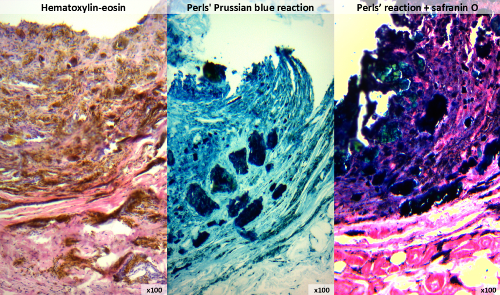
A biopsy of the skin of the finger of an 84-year-old patient with clinical suspicion of melanoma was sent to the histology laboratory. Macroscopically, there was a 2×2 mm area of pigmentation on the skin surface, and a black pigment was visualised on the section. During histological examination, the pathologists suspected that this was not melanoma at all, but rather hemosiderin deposits from an old haemorrhage. But suspicion is not enough, because the patient’s further fate can vary from complete recovery to amputation… Therefore, it was decided to use Perl’s reaction to detect hemosiderin. During this staining, the tissue is treated with solutions of yellow blood salt and hydrochloric acid, as a result of which iron compounds become blue in colour – Prussian blue is formed (yes, the same one you heard about in school chemistry lessons). The first photo shows haematoxylin and eosin staining; the second shows a positive Perls’ reaction; and the third shows additional nuclei stained with safranin O. Now the pathologists were sure this was an old haemorrhage, not melanoma. We express our sincere gratitude to the head of the Department of Theoretical and Applied Chemistry, Lyudmila Ponomaryova, for her help with the reagents!